Home » Services » FISH Testing
FISH Testing
Fluorescent in situ hybridization, commonly known as FISH testing, allows pathologists to detect specific genetic abnormalities directly within tissue samples. Golden State Dermatopathology provides advanced FISH testing to support more accurate diagnoses and help guide treatment decisions when standard lab results need additional confirmation.
How Does FISH Testing Work?
FISH testing uses fluorescent probes that attach to specific genes or chromosomes within tissue samples. When these probes find their target, they light up under a special microscope, allowing pathologists to see genetic changes that cause cancer or other diseases.
This technique is important because it can detect genetic abnormalities that regular microscopy cannot see. While traditional tissue examination looks at cell shape and structure, FISH testing reveals the underlying genetic changes that drive disease development.
Golden State Dermatopathology uses advanced FISH testing to provide definitive answers when standard tissue examination alone cannot determine whether a lesion is benign or malignant. This molecular evaluation transforms uncertain diagnoses into clear results that guide appropriate treatment decisions.
Why FISH Testing Matters
The integration of FISH testing into routine dermatopathological practice offers several advantages that improve diagnostic confidence and patient care. Increased diagnostic accuracy is the primary benefit, as FISH testing provides objective molecular evidence that supports or refines morphological impressions.
Speed of results makes FISH testing particularly beneficial compared to other techniques. While comprehensive genetic sequencing may require days or weeks, FISH testing typically provides results within 24-48 hours, allowing for timely clinical decision-making without significant delays in patient care.
Tissue preservation requirements for FISH testing align well with routine processing. The same tissue blocks used for standard H&E staining can serve as the source material for FISH analysis, eliminating the need for special tissue collection protocols.
The FISH Testing Process
At Golden State Dermatopathology, the FISH testing process is carefully structured to ensure accurate, high-quality results.
It begins with an initial review of the case to determine whether FISH testing is appropriate, based on what the tissue looks like under the microscope, the patient’s medical history, and any unanswered diagnostic questions.
Next, pathologists select the most relevant area of the biopsy sample, focusing on the parts most likely to contain the information needed. They closely examine stained tissue sections and mark the exact spots where FISH testing will be most helpful.
The type of FISH probe used depends on the clinical concern and what condition is being considered. Different probes are designed to detect specific genetic changes linked to certain types of tumors, and the chosen panel reflects the possibilities being investigated.
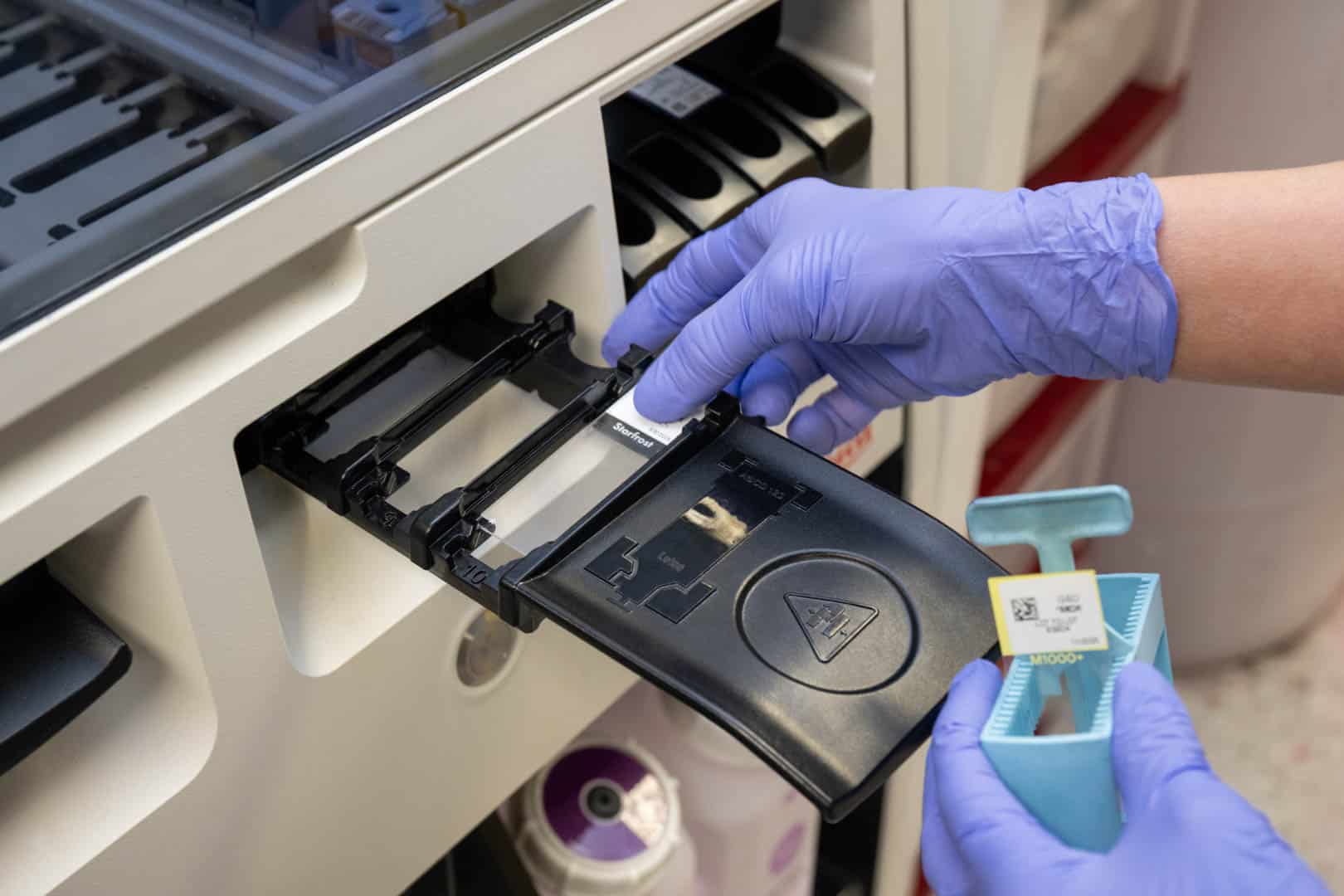
The actual testing requires precise handling, including careful control of temperature, timing, and chemical steps, to ensure the probes attach correctly to their genetic targets. Each step is monitored through strict quality control to make sure the results are reliable.
Finally, skilled dermatopathologists interpret the fluorescent signals, looking at how they appear, where they are located, and how strong they are. They compare these patterns to established guidelines to identify whether any significant genetic abnormalities are present.
Advancing Dermatopathological Excellence Through Molecular Precision
FISH testing plays an important role in modern skin pathology by adding clear, molecular-level evidence to help improve diagnostic accuracy.
At Golden State Dermatopathology, advanced FISH testing is paired with expert analysis of tissue samples to deliver thorough and reliable diagnoses.
This combination supports better patient care and positions the practice as a trusted resource for complex cases that require deeper diagnostic insight.


